
Description
PrymaLab · Research Use Only
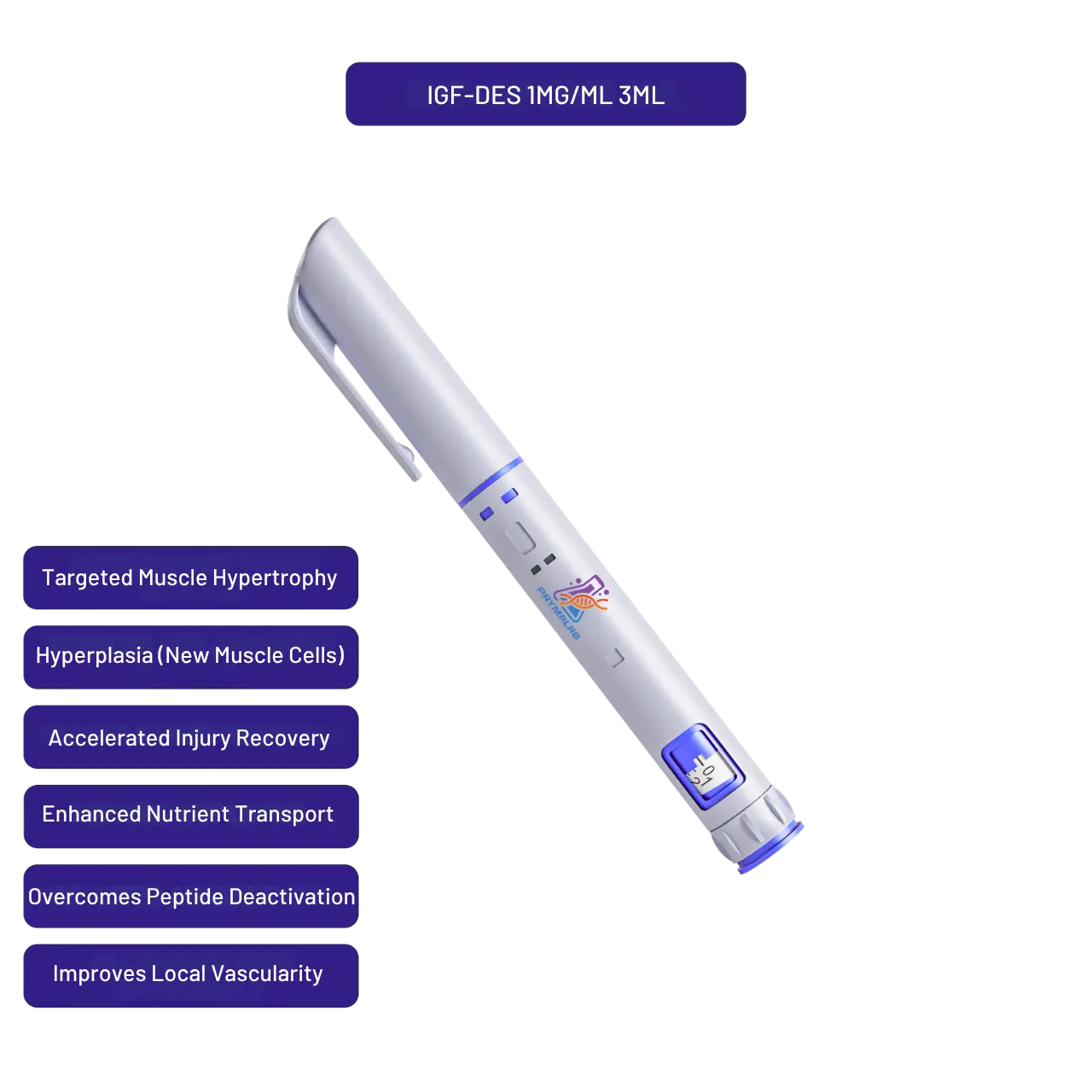
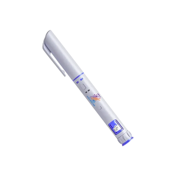
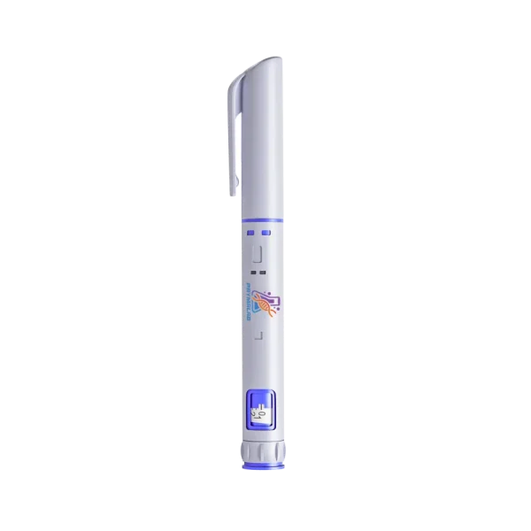
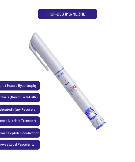
IGF-1 DES Autoinjector
DES(1-3) IGF-1 · 67 residues · pre-filled device
IGF-1 DES autoinjector is a pre-filled device supplying DES(1-3) IGF-1, a 67-residue truncated analogue of insulin-like growth factor 1. The designation indicates removal of the first three residues, glycine, proline and glutamate, from the amino terminus of the 70-residue native sequence.
Specification Table
| Property | Value |
|---|---|
| Compound | DES(1-3) IGF-1 |
| Common designation | IGF-1 DES |
| Parent molecule | Insulin-like growth factor 1, 70 residues |
| Residue count | 67 |
| Modification | Removal of residues 1 to 3 from the amino terminus |
| Residues removed | Glycine-Proline-Glutamate (GPE) |
| Approximate molecular weight | 7371 Da |
| Disulfide bonds | Three, as in native IGF-1 |
| Molecular target | IGF-1 receptor (IGF1R), a receptor tyrosine kinase |
| Effect of truncation | Substantially reduced affinity for IGF binding proteins |
| Natural occurrence | Reported as a naturally occurring form in some tissues |
| Relationship to GPE | The removed tripeptide has been studied separately as a distinct compound |
| Format | Pre-filled autoinjector device |
| Physical state | Solution, supplied ready-filled |
| Appearance | Clear colourless solution |
| Purity | Per lot-specific certificate of analysis |
| Storage | 2-8°C, protected from light. Do not freeze the device |
| Regulatory status | No approved human or veterinary formulation in any jurisdiction |
Why Does Removing Three Residues Change So Much?
The IGF-1 DES autoinjector supplies a protein missing three residues from seventy is a small change by mass and a large one by function, which makes the reason worth examining.
The amino-terminal region of IGF-1 participates directly in the binding protein interface. Glutamate at position 3 in particular contributes an acidic contact that IGF binding proteins recognise, and removing it eliminates that contact rather than merely weakening it.
Note the convergence with the LR3 analogue, which modifies the same position by substitution rather than deletion. Two different chemical strategies aimed at the same residue, arrived at independently, which is reasonable evidence that position 3 genuinely matters for binding protein association.
Receptor binding is comparatively less affected, because the receptor contact surface involves different regions of the molecule. The truncated protein retains the three disulfide bonds and the overall fold that receptor binding depends on.
The removed tripeptide, glycine-proline-glutamate, has been studied as a compound in its own right, which is an unusual situation where both the fragment and the remainder attract separate research attention.
How Do DES and LR3 Differ as Research Tools?
Both analogues address binding protein affinity, and choosing between them depends on what a design needs to hold constant.
DES(1-3) is smaller than native IGF-1, at 67 residues against 70, while LR3 is larger at 83, because it adds an extension as well as substituting. Molecular weight differs accordingly, roughly 7371 against roughly 9111 daltons.
That weight difference matters for molar calculation. Two preparations at the same mass concentration contain substantially different molar concentrations, and comparing analogues on a mass basis rather than a molar basis introduces an error of over twenty percent.
DES(1-3) is reported to occur naturally in some tissues, whereas LR3 is entirely synthetic with no natural counterpart. For work concerned with physiological relevance that distinction may matter.
LR3 carries a larger structural alteration, with thirteen added residues representing a substantial addition to a small protein, and whether that extension affects anything beyond binding protein association is a question a DES comparison can help answer.
Running both analogues in parallel is more informative than either alone where the question concerns binding protein contribution.
What Happens to Folded Proteins in Solution?
A pre-filled device holds this protein in solution continuously, and the degradation routes available to a folded protein differ from those affecting short peptides.
Aggregation is the principal concern. Partially unfolded molecules expose hydrophobic surfaces that normally sit buried in the core, and those surfaces associate with each other. The process is self-accelerating, since aggregates nucleate further aggregation, and it is effectively irreversible.
Disulfide scrambling is the second route. Three disulfide bonds define the fold, and under circumstances permitting thiol-disulfide exchange those bonds can reform in incorrect pairings. The rearranged molecule has the same mass and a different structure, which makes the change invisible to mass spectrometry.
Surface adsorption removes protein from solution onto container walls, disproportionately at low concentration, and deamidation and oxidation also proceed, though more slowly than aggregation under typical storage.
Air-liquid interfaces accelerate unfolding, which is why agitation is specifically discouraged. Every bubble generated presents a surface at which protein molecules unfold.
What Should Be Verified Before Using the Device?
A sealed solution-state protein device limits verification, and knowing what remains available is worth establishing before the material is needed.
Visual inspection carries real information here, more than for a small molecule. The solution should be clear and colourless. Cloudiness, opalescence or visible particulate indicates aggregation and disqualifies the unit, since aggregated protein has lost the fold determining its activity.
The certificate of analysis should state protein concentration determined analytically, purity by a method appropriate to proteins such as size-exclusion chromatography, and the absence of aggregate. Size-exclusion is specifically useful because it detects soluble aggregates that visual inspection cannot.
Manufacture date matters for the same reason it matters for solution-state NAD+, though the mechanism differs. Aggregation accumulates with time in solution, and a recently manufactured device has had less opportunity for it.
Storage history during transit should be documented. A cold-chain excursion is more consequential for a folded protein than for most compounds, because that aggregation does not reverse when temperature is restored.
What Is GPE and Why Does the IGF-1 DES Autoinjector Question Involve It?
The three residues removed to produce this analogue have an independent research history, which is unusual and worth knowing about.
Glycine-proline-glutamate, abbreviated GPE, is the amino-terminal tripeptide of IGF-1. When the parent protein is cleaved to produce the truncated form, the tripeptide is released as a separate molecule rather than degraded immediately.
GPE has been studied in its own right, principally in neural contexts, and reported not to act through the IGF-1 receptor. Its proposed interactions involve other systems entirely, which means the cleavage produces two molecules with different targets rather than one active molecule and one waste product.
The experimental implication is direct. In any system where the parent protein is subject to amino-terminal cleavage, both products are present, and an observed effect could originate from either.
Supplying the truncated form removes that ambiguity from the design. The tripeptide is simply absent, so anything observed is attributable to the 67-residue species.
Conversely, comparing the truncated form against the intact parent in a cleaving system does not isolate the truncation, because the parent condition also generates the tripeptide. Running GPE as a third arm resolves it.
How Do the Three Disulfides Constrain Handling?
The disulfide architecture is what makes this a folded protein rather than a flexible chain, and it sets several handling boundaries.
Three bonds in a 67-residue protein is a high density, and they cross-link the chain into a compact fold. The insulin-like architecture is stabilised almost entirely by these bonds rather than by an extensive hydrophobic core.
Reducing agents are therefore excluded. Dithiothreitol, beta-mercaptoethanol and tris(2-carboxyethyl)phosphine will break the bonds, and the protein does not refold correctly afterward without controlled oxidative conditions.
That matters because reducing agents appear routinely in buffers for other purposes, including protease inhibition and protection of unrelated proteins in a lysate. A buffer chosen for a downstream step can inactivate the growth factor before it acts.
Free thiols in the system present the same hazard through disulfide exchange, which can occur even without a formal reducing agent present when cysteine-containing proteins are abundant.
Checking buffer composition for reducing agents before adding this protein is a small step that spares an entire experiment from a false negative.
What Should an IGF-1 DES Autoinjector Record Include?
Documentation for a solution-state protein carries one field that dominates all the others.
Storage history, including every temperature excursion and its duration. Aggregation is irreversible, so an excursion three weeks ago still affects the material today, and no subsequent handling undoes it.
Beyond that, the standard fields apply: lot number, fill date, measured protein concentration with the method used, and purity by size-exclusion chromatography rather than by a method blind to soluble aggregate.
Visual condition at each use is worth logging rather than merely checking. A device that was clear on Monday and faintly hazy on Friday has told you something about its storage that a single pass-fail check would discard.
Where an IGF-1 DES autoinjector is used across a series, note which experiments drew from which unit.
Storage and Handling in Laboratory Practice
An IGF-1 DES autoinjector wants cold, dark, still and unfrozen storage, inspected each time.
Maintain 2-8°C without interruption. Aggregation rate rises with temperature and the process does not reverse, so an ambient excursion has permanent consequences rather than temporary ones.
Never freeze. Ice formation concentrates solutes in the shrinking liquid phase and creates ice-water interfaces, and both drive aggregation efficiently. This applies to the device format regardless of how a bulk protein solution might be handled with cryoprotectant.
Handle gently. Do not shake, do not invert repeatedly, and avoid generating bubbles, because air-liquid interfaces unfold protein and mechanical agitation creates them.
Inspect for clarity before every use and reject any unit showing cloudiness or particulate. Record lot, manufacture date, storage history including any excursion, and dates of use.
One further point concerns temperature during use rather than during storage. A device removed from refrigeration warms quickly, and aggregation rate rises with temperature.
Where a session involves several sequential measurements, returning the device to cold between them rather than leaving it on the bench is worth the small inconvenience. The alternative is an hour at ambient temperature per session, accumulating across a study.
That accumulated warm time is rarely recorded and it is a plausible explanation for drift in a long series.
What Does the IGF-I System Actually Consist Of?
The endocrinology here is well established, and understanding it explains both why this analogue exists and why the claims around it outrun the evidence.
IGF-I, historically called somatomedin C, is a 70-residue protein produced largely by the liver in response to growth hormone.
Most of the growth-promoting activity attributed to growth hormone is actually mediated by IGF-I downstream of it, which is why the two are discussed together.
Almost none of it circulates free. IGFBPs, the six binding proteins, hold the great majority in complex, with IGFBP-3 carrying most of the circulating pool.
That IGFBP binding is what sets the half-life. Bound IGF-I persists for hours; free IGF-I is cleared in minutes.
Signalling runs through the IGF-1 receptor and then down the PI3K/Akt/mTOR pathway, which is the canonical route to increased protein synthesis in muscle.
Reported downstream effects in that literature include satellite cell activation, muscle protein synthesis, collagen synthesis and glucose uptake into tissue.
Because the receptor is closely related to the insulin receptor, IGF-I also lowers blood glucose, and that cross-reactivity is the source of the safety concern discussed below.
An approved IGF-1 product exists. Increlex, mecasermin, is recombinant human IGF-I approved for severe primary IGF-1 deficiency, and its label is where the real safety data for this class lives.
How Do DES, LR3 and MGF Differ?
Three analogues circulate in this market and the differences between them are entirely about escaping the binding proteins.
This compound removes the first three N-terminal amino acids from native IGF-1, which is the region the binding proteins engage. Removing it drops IGFBP binding by more than an order of magnitude.
The consequence is higher biological activity at the receptor in the presence of binding proteins, with receptor affinity for the IGF-1 receptor itself broadly preserved.
IGF-1 LR3 takes a different route to the same destination, adding a 13-residue extension and substituting one residue, which also reduces IGFBP binding while extending the half-life considerably.
So DES is short-acting and locally biased; IGF-1 LR3 is long-acting and systemic. That is the practical distinction between them and it is a pharmacokinetic one rather than a potency one.
MGF, mechano growth factor, is a splice variant of IGF-1 expressed after mechanical loading, and it is a third thing again rather than a variant of either. The MGF sold in this market is usually a short synthetic peptide from its C-terminal region rather than the full splice variant.
That distinction matters more than it sounds. A 24-residue peptide and a 67-residue folded protein have different handling requirements, different stability profiles and different analytical needs.
This compound is a protein rather than a peptide in the strict sense, and it is sold in a market organised around peptide products, which is why the two words get used interchangeably for it.
Anyone reading results across the three is reading across different pharmacokinetic profiles, so a finding for one does not transfer.
What Is the Safety and Anti-Doping Position?
Two things need stating plainly and neither appears often enough in listings for this compound.
The first is hypoglycemia. Because the IGF-1 receptor and the insulin receptor are related, this class lowers blood glucose, and low blood sugar is the dose-limiting adverse effect on the Increlex label.
Reported side effects for the approved product also include injection site reactions and tonsillar hypertrophy, with hypoglycemia serious enough that the label directs dosing with food. Lipohypertrophy at a repeatedly used injection site is the other documented local effect, and tissue repair at the site is slower than the market assumes.
A research analogue engineered specifically to evade the binding proteins that would normally buffer it is, by design, less buffered against that effect than native IGF-1 is.
The second is anti-doping. The World Anti-Doping Agency prohibits IGF-1 and its analogues, and they sit on the WADA prohibited list in and out of competition under the growth factor category.
Any tested athlete using this compound is committing an anti-doping rule violation whatever the format or the label, and that is worth knowing before rather than after.
The theoretical concern raised most often is that IGF-1 signalling promotes cell proliferation broadly, so sustained elevation is discussed in relation to tumour growth. No study has examined that for this analogue over any meaningful period.
This device is supplied for laboratory research only and is not for administration to humans or animals.
What Do the Circulating Protocols Represent?
Usage guidance for this compound circulates widely, and its origin is worth understanding before any of it is repeated.
Figures are quoted in mcg rather than milligrams, reflecting the potency of the class, usually with a post-workout timing and sometimes a pre-workout one.
The rationale offered is that a short half-life suits administration near resistance training, when muscle stem cells and satellite cells are already activated by the mechanical stimulus.
That is a plausible-sounding argument built on no human pharmacokinetic profile for this analogue, and no published study establishes a dosing interval for it.
Cycling protocols and dosing protocols for it come from forums and vendor sheets, and receptor downregulation is invoked as the reason for cycling without measurement supporting it.
Intramuscular injection is promoted on the theory that local delivery concentrates the effect in the trained muscle, which is the reasoning behind the site-injection practice generally.
Subcutaneous injection, sometimes written subq, is the alternative, and no comparison of the two has been published for this compound.
Claims about muscle growth, muscle hypertrophy, hyperplasia, anabolism, nutrient uptake and water retention all come from that same market rather than from trials.
Hypertrophy and hyperplasia are worth distinguishing, since the first means larger existing fibres and the second means more of them, and only the first is established for this class in humans.
The compound is stacked with CJC-1295, ipamorelin and other GH secretagogues, with SARMs and anabolic steroids in some guides, and with BPC-157 for injury recovery or injury rehabilitation.
CJC-1295 and ipamorelin work upstream, prompting growth hormone release so that IGF-I rises indirectly. This peptide is the downstream product itself, which is why stacking both is described as redundant by some of the same guides that recommend it.
Angiogenesis and improved insulin sensitivity are attributed to the class in places, the first from IGF-I’s role in vascular growth and the second from its insulin-receptor cross-reactivity, and neither has been measured for this analogue.
Doses quoted in mcg per site, several times weekly, are the usual pattern. A research peptide sold in vials at unstated concentration makes even those figures unverifiable, since a mcg figure requires knowing the concentration it came from.
Anabolism in this context means the whole set of building processes the pathway drives, and it is a description of a mechanism rather than a measured outcome.
Oral bioavailability for a 67-residue protein is effectively zero, which is why no oral format of this compound exists despite the market’s appetite for one.
None of those combinations has been tested. Bovine colostrum appears in the same guides as a supposed natural source, which it is not in any meaningful quantity.
Muscle wasting and age-related muscle loss are real clinical problems, and Increlex rather than a research chemical is where the approved evidence for this class sits.
Published Literature
References verified against the publisher record. The IGF-1 analogue literature is well established and independent.
- Ballard FJ, Francis GL, Ross M, Bagley CJ, May B, Wallace JC. Biochemical and Biophysical Research Communications. 1987;149(2):398-404.
- Sara VR, Carlsson-Skwirut C, Drakenberg K, Giacobini MB, Ho¨kfelt T, Schwartz PH. Annals of the New York Academy of Sciences. 1993;692:183-191.
- Baxter RC. Nature Reviews Endocrinology. 2014;10(4):201-212.
- Firth SM, Baxter RC. Endocrine Reviews. 2002;23(6):824-854.
- Francis GL, Ross M, Ballard FJ, Milner SJ, Senn C, McNeil KA, Wallace JC, King R, Wells JR. Journal of Molecular Endocrinology. 1992;8(3):213-223.
Frequently Asked Questions
What is the IGF-1 DES autoinjector?
A pre-filled device supplying DES(1-3) IGF-1, a 67-residue truncated analogue lacking the first three residues of the native 70-residue sequence. Those residues are glycine, proline and glutamate. It is supplied as a laboratory research reagent only, with no human or veterinary use.
Why does removing three residues matter so much?
Because the amino-terminal region participates directly in the binding protein interface. Glutamate at position 3 contributes an acidic contact that IGF binding proteins recognise, and removing it eliminates that contact rather than merely weakening it.
Is there a connection to the LR3 analogue?
Yes, and an informative one. LR3 modifies the same position by substitution rather than deletion. Two different chemical strategies aimed at the same residue, arrived at independently, is reasonable evidence that position 3 genuinely matters for binding protein association.
Does the truncation affect receptor binding?
Comparatively less, because the receptor contact surface involves different regions of the molecule. The truncated protein retains the three disulfide bonds and the overall fold that receptor binding depends on.
How do DES and LR3 compare by size?
DES(1-3) is 67 residues at roughly 7371 daltons. LR3 is 83 residues at roughly 9111 daltons. That difference means two preparations at the same mass concentration contain substantially different molar concentrations, an error of over twenty percent if compared by mass.
Does either occur naturally?
DES(1-3) is reported as a naturally occurring form in some tissues. LR3 is entirely synthetic with no natural counterpart, which is a distinction that may matter for work concerned with physiological relevance.
Why run both analogues in parallel?
Because LR3 carries a larger structural alteration, with thirteen added residues representing a substantial addition to a small protein, and whether that extension affects anything beyond binding protein association is a question a DES comparison helps answer.
What degradation routes affect a folded protein?
Aggregation principally, where partially unfolded molecules expose buried hydrophobic surfaces that associate with each other. The process is self-accelerating and effectively irreversible. Disulfide scrambling, surface adsorption, deamidation and oxidation also occur.
Why is disulfide scrambling hard to detect?
Because the rearranged molecule has the same mass as the correctly folded one, only with bonds reformed in incorrect pairings, and mass spectrometry cannot distinguish them, so detection requires methods sensitive to structure rather than to mass.
What should the certificate state?
Protein concentration determined analytically, purity by a protein-appropriate method such as size-exclusion chromatography, and absence of aggregate. Size-exclusion specifically detects soluble aggregates that visual inspection cannot see.
What is IGF-1 DES?
Insulin-like growth factor 1 DES(1-3), also written IGF-1 DES1-3 or IGF-DES, is native IGF-1 with the first three N-terminal amino acids removed. That region is what the binding proteins engage, so removing it drops IGFBP binding by more than an order of magnitude.
How does it differ from IGF-1 LR3?
Both evade the binding proteins by different routes. IGF-1 LR3 adds a 13-residue extension and substitutes one residue, giving a much longer half-life. This one is short-acting and locally biased. The difference is pharmacokinetic, not a matter of potency.
Is it banned by WADA?
Yes. IGF-1 and its analogues sit on the WADA prohibited list in and out of competition under the growth factor category. A tested athlete using this compound is committing an anti-doping rule violation whatever the format or label.
What is the main safety concern?
Hypoglycemia. The IGF-1 receptor is closely related to the insulin receptor, so this class lowers blood glucose, and low blood sugar is the dose-limiting effect on the Increlex label. An analogue engineered to evade the binding proteins is less buffered against it than native IGF-1.
Where do the mcg dosing figures come from?
Forums and vendor sheets. No human pharmacokinetic profile for this analogue has been published, so no dosing interval can be derived from it. Cycling protocols invoke receptor downregulation as a rationale without measurement behind it.
Does it need reconstitution?
Not in this format, which ships in solution. Research peptide and protein products sold in vials as lyophilized powder need bacteriostatic water and a reconstitution step, and for a folded protein like this one that step also risks aggregation.
Does it build muscle?
The IGF-I system drives muscle protein synthesis through the PI3K/Akt/mTOR pathway, and that biology is established. Whether administering this analogue produces muscle growth in humans has not been tested in a controlled trial, and claims about tissue repair and cellular regeneration come from the same place.
How should a supplier verify identity?
Mass confirmation against the expected value plus HPLC purity with the gradient stated, and size-exclusion for aggregate. For peptide hormones of this size the folded state matters as much as the sequence, which a purity percentage alone does not report.
Compliance Statement
IGF-1 DES is sold exclusively for laboratory research use. It is not a drug, food, or cosmetic product, and it is not a dietary product of any kind. It is not approved by the FDA or any comparable authority for human or veterinary use, and the pre-filled device format does not alter that status in any way. This product is not intended to diagnose, treat, cure, or prevent any disease. It must not be given to humans or animals. Purchase is restricted to qualified researchers and institutions operating within applicable laws. All handling is the responsibility of the purchasing laboratory.











12 reviews for Preloaded Autoinjector | IGF-DES | 3ml Pen | 1mg/ml